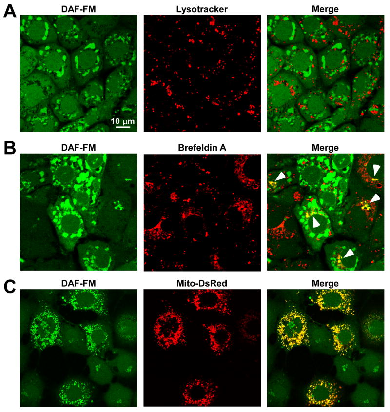
Figure 3.
Subcellular localization of DAF-NO adduct following NO delivery. DAF-loaded RPMVECs exposed to DeaNONOate (5 μM) were stained with (A) Lysotracker Red and (B) brefeldin A- BODIPY 558/568 conjugate to simultaneously assess DAF-NO adduct localization in the lysosomes and golgi apparatus, respectively. (C) RPMVECs transfected with mitochondrially-targeted DsRed (Mito-DsRed) were also loaded with DAF to assess DAF-NO adduct localization in the mitochondria following DeaNONOate application.

