Figure 4.
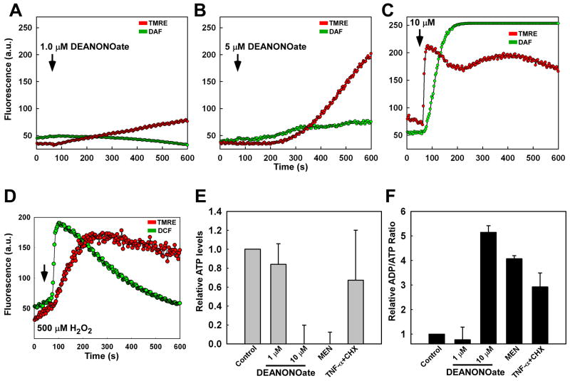
NO alters mitochondrial function and bioenergetics in endothelial cells. RPMVECs were simultaneously stained with DAF-FM and the mitochondrial membrane potential indicator dye tetramethyl-rhodamine-ethyl ester (TMRE; 50 nM) and imaged continuously via confocal microscopy. Mean representative traces of DAF and TMRE fluorescence were quantitated following application of (A) 1.0 μM, (B) 5.0 μM, and (C) 10.0 μM DeaNONOate. (D) The kinetics of hydrogen peroxide (H2O2)-induced fluorescence (CMH2DCF-DA) and mitochondrial membrane potential alterations. (E) ATP levels were assessed 12 hr following NO addition and normalized to untreated RPMVECs. (F) Ratio of ADP/ATP in RPMVECs. Menadione (20 μM) and TNF-α (20 ng/ml) + cycloheximide (CHX; 1 μM) were used as positive controls for necrosis and apoptosis, respectively.

