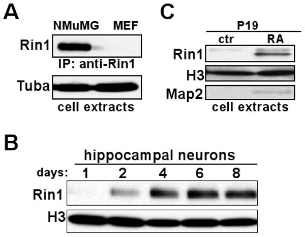
Figure 1.

Restricted Expression of Rin1. A. Immunoprecipitation and immunoblot of cell extracts from NMuMG and primary MEF cells using anti-Rin1 (polyclonal and monoclonal, respectively). Total protein used for immunoprecipitation was normalized using Bradford assay and confirmed with an anti-α-tubulin (Tuba) blot. B. Immunoblot of extracts from mouse hippocampal neurons grown in culture for the indicated time, using anti-Rin1 and normalized using anti-histone 3 (H3). Histone expression was used because tubulin levels change during this period of extensive neurite outgrowth. C. Immunoblot of extracts from P19 cells grown under control conditions (ctr) or after neuronal differentiation with retinoic acid (RA). The anti-Rin1 signal was normalized using anti-H3, and anti-Map2 was used to validate differentiation.
