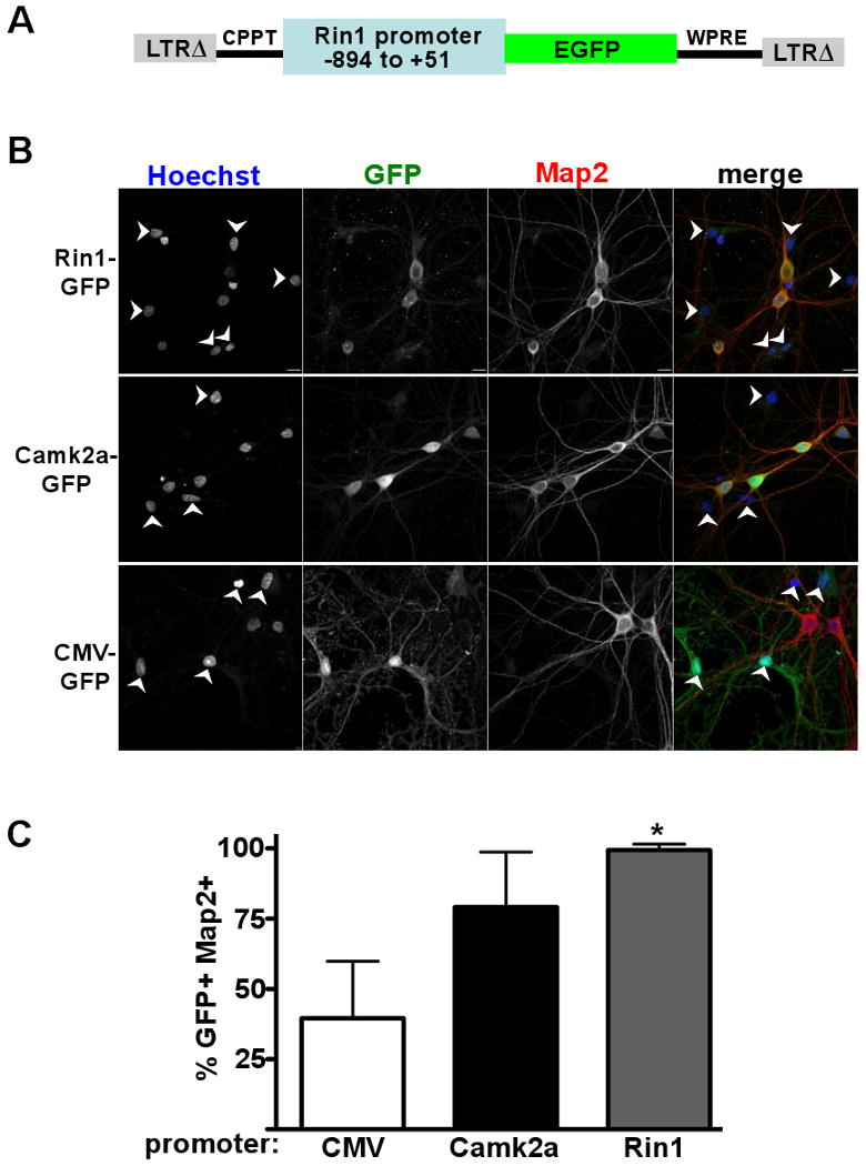
Figure 2.

A Rin1 promoter fragment confers neuronal expression to a GFP reporter. A. Diagram of the lentivirus construct used in the expression experiments. LTRΔ = long terminal repeat with self-inactivating deletion; CPPT = central polypurine tract; GFP = green fluorescent protein; WPRE = woodchuck hepatitis virus post-transcriptional regulatory element. B. Rat hippocampal neurons infected with the promoter-GFP virus indicated at left, and visualized as indicated above with Hoechst (nuclear stain), anti-GFP, anti-Map2 and as a merged image. Arrows indicate cells not stained with anti-Map2. Scale bars (10 microns) are in bottom right of top row images. C. Statistical analysis of infected cultures shown in B. For each cell population, 12 independent fields (> 450 total cells) were evaluated. GFP and Map2 double immunofluorescent cells (GFP+ and Map2+) are given as a percentage of total GFP+ cells, with standard deviation. * p < 0.001 (comparing Rin1 and CMV promoter signals)
