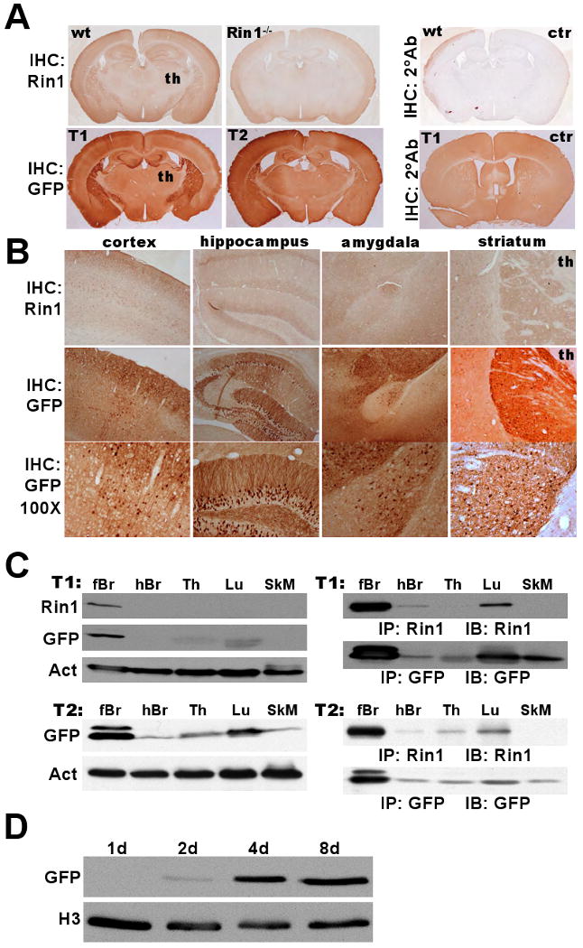
Figure 3.

Rin1 promoter-driven GFP expression in transgenic mice. A. Top: Wild type (wt) or Rin1-/- mouse brain coronal sections subjected to immunohistochemistry (IHC) staining with anti-Rin1 or secondary antibody (2°) alone as control (ctr). Signal in thalamus (th) is near background level. Bottom: Two independently derived Rp-GFP transgenic animals (T1 & T2) stained with anti-GFP or secondary antibody alone (ctr). B. Top: Rin1 staining in designated telencephalic regions of wild type brain (th = thalamus). Middle: GFP staining from T1 tissue (40× mag). Bottom: same as above (100× mag). C. Immunoblot detection of Rin1 and GFP expression in T1 and T2 transgenic mouse tissues (forebrain (fBr), hindbrain (hBr), thymus (Th), lung (Lu) and skeletal muscle (SkM)). Left: Direct immunoblot of tissue extracts, with actin (Act) normalization. Right: Immunoblot (IB) of immunoprecipitated (IP) material from tissue extracts. D. Dissociated hippocampal neurons from a transgenic (T1) mouse cultured for the indicated time and immunoblotted with anti-GFP, normalized with anti-histone 3 (H3).
