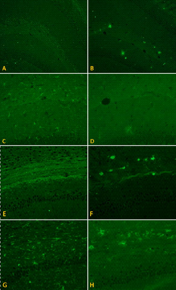
Figure 4. Immunohistochemical staining of amyloid plaques and microglia.

Coronal paraformaldehyde-fixed paraffin embedded sections were labeled with antibodies against Aβ(6E10) and ionized calcium binding protein 1 (Iba 1), a marker for microglia. In the molecular layer of the hippocampus just superior to the dentate gyrus in the M631L mice (A) there were few plaques but distinctive punctate staining; in the J20 (B), however, greater plaque formation was seen in the same area (6E10 images at 10x), but no punctate staining. The punctate pattern of Aβ distribution was associated with microglial activation in the M631L (C), which was not observed in the J20, the latter of which instead showed amoeboid phagocytic microglia surrounding plaques (D) (Iba 1 images at 20X). The punctate labeling in the M631L mice was even more marked in the corpus callosum (E) and the white matter tracts lateral to the hippocampus and surrounding the thalamus, associated with a proportionately greater microglial response (G); again, the J20 did not demonstrate punctate staining, but instead formed plaques in the corpus callosum (F), surrounded by phagocytic microglia (H) (all 20X).
