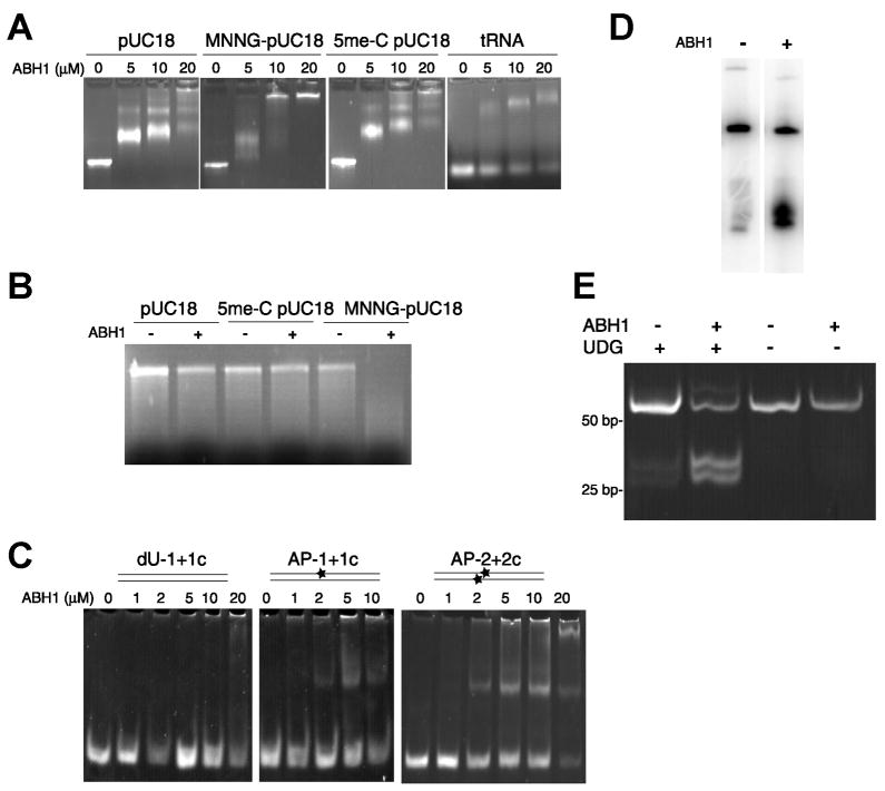
Fig. 1.
Binding and cleavage of DNA by ABH1. (A) ABH1 binds to DNA and RNA. Linearized pUC18 (0.3 – 0.5 μg) was used directly, treated with MNNG to produce predominantly 7-meG and 3-meA, or subjected to SssI methyltransferase and S-adenosyl methionine to generate 5-meC-containing DNA. ABH1 was mixed with these samples, electrophoresed on 1% agarose, and stained with EtBr. (B) Degradation of MNNG-treated DNA by ABH1. Unmodified, SssI-methylated, and MNNG-treated linearized pUC18 (appr. 0.5 μg) were incubated with ABH1 (10 μM) for 3 h at 37 °C, electrophoresed, and stained. (C) ABH1 binding to dU- and AP-containing oligonucleotides. ABH1 was mixed with 100 ng of (i) oligo1 (dU at position 28) annealed to the complementary oligo1c, (ii) oligo1+oligo1c treated with UDG (30 min at 37 °C) to generate an abasic site (depicted as a star), and (iii) oligo2+oligo2c (both with dU) treated with UDG. The samples were electrophoretically analyzed. (D) ABH1 cleavage of oligonucleotides containing an abasic site. Oligo1 labeled with 32P at its 5′ end was annealed to oligo1c and treated with UDG. The effect of ABH1 (1 μM, 1 h at 37 °C) on DNA cleavage was examined by gel electrophoresis. (E) ABH1 cleavage of ds-DNA. ABH1 (5 μM) was incubated at 37 °C with oligo2 annealed to oligo2c (0.3 μg) containing either dUs or abasic sites in close proximity on opposed strands. After 90 min, samples were incubated with proteinase K at 65 °C for 1 h and analyzed by electrophoresis.

