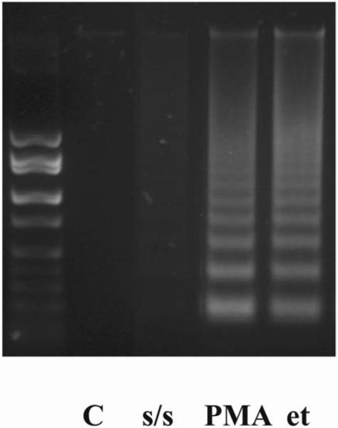
Figure 2. DNA fragmentation in thymocytes after PMA, serum starvation and etoposide treatment.
Cells were kept under different conditions (1 μM PMA or 100 μM etoposide), including serum starvation, for 4 hours. DNA fragmentation was promoted in PMA and etoposide treated thymocytes in comparison with serum starved cells. Cells exposed to vehicle (DMSO) did not differ from untreated cells.

