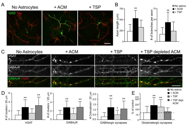
Figure 4. TSPs do not increase GABAergic axon length, branching, or synaptogenesis.
Hippocampal neurons were cultured alone, with ACM, with TSP-1, or with TSP-depleted ACM, and were immunostained at 7 div with antibodies against GAD (green) and tau (red) to label GABAergic axons. (A), GABAergic axon length and branching were increased in neurons cultured with ACM (middle) but not when neurons were treated with TSP-1 (right) compared to neurons cultured alone (left) at 4 div. Scale bar = 25 μm. (B), Quantification shows that GABAergic axon length and branching were significantly increased in neurons cultured with ACM compared to neurons treated with TSP-1 and neurons cultured alone. All values are shown as mean ± s.d. (n = 133 cells, 4 independent expts.; Kruskal-Wallis nonparametric ANOVA test followed by Dunn’s pairwise multiple comparison test, p < 0.001). (C), Hippocampal neurons were cultured alone, with ACM, with TSP-1, or with TSP-depleted ACM, and were immunostained at 7 div with antibodies against VGAT (red), and GABAAR β3 subunit (green) to label GABAergic synapses. An increase in presynaptic VGAT clusters, postsynaptic GABAAR clusters and GABAergic synapse density were observed at 7 div in neurons cultured with ACM (middle left) or with TSP-depleted ACM (right) but not when neurons were treated with TSP-1 (middle right) compared to neurons cultured alone (left). Scale bar = 10 μm. (D), Quantification of increase in presynaptic VGAT clusters (left), postsynaptic GABAAR clusters (middle) and GABAergic synapse density (right) per length dendrite in neurons treated with ACM, TSP-1, or TSP-depleted ACM. All values are shown as mean ± s.d. (n = 75 cells, 4 independent expts.; Kruskal-Wallis nonparametric ANOVA test followed by Dunn’s pairwise multiple comparison test, Single asterisk indicates p < 0.05, ** indicates p < 0.001). (E), Quantification of increase in glutamatergic synapse number per dendrite length (clusters double stained with VGlut and PSD-95 antibodies) in neurons treated with ACM, TSP-1 but not TSP-depleted ACM. TSPs increase glutamatergic synapse number (see also Christopherson et al., 2005). All values are shown as mean ± s.d. (n = 59 cells, 4 independent expts.; Kruskal-Wallis nonparametric ANOVA test followed by Dunn’s pairwise multiple comparison test, p < 0.001).

