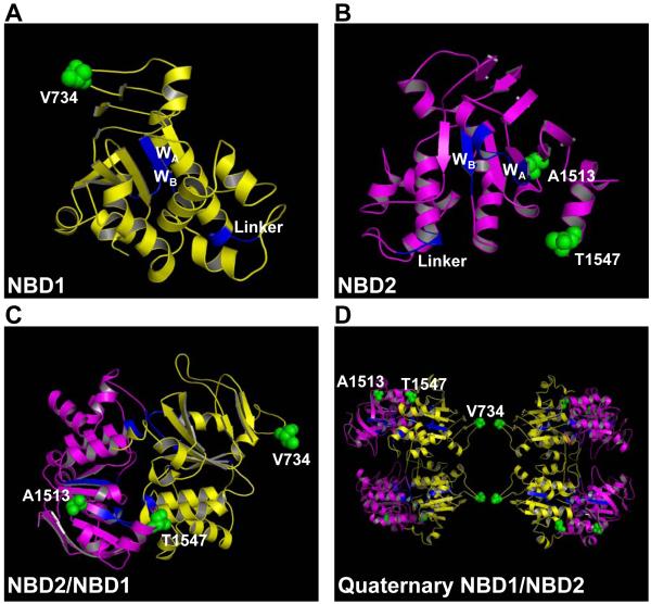
Figure 5.
Structure of NBDs incorporating pertinent pathogenic mutations found in human disease. (A) Homology structural model of NBD1 (A), NBD2 (B), NBD1/NBD2 (C), and SAXS resolved quaternary structure of NBD1/NBD2 (D) with mutated residues clustered at protein-protein interaction locales. NBD1 in yellow; NBD2 in purple; Walker A (WA), Walker B (WB), and linker motifs in blue; pathogenic human mutations in green.

