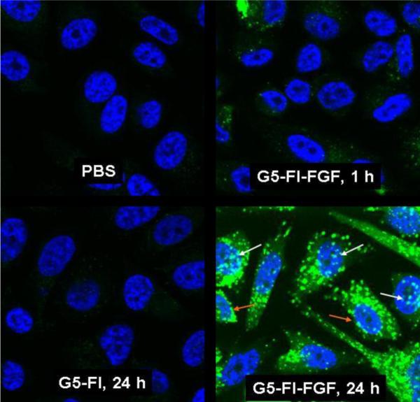
Figure 5.

Internalization of G5-FI-FGF in PC3 cells. Cells grown on cover slips were incubated with 300 nM each of G5-FI-FGF or the control conjugate G5-FI at 37°C for 1 or 24 hours. Cells were rinsed and fixed with paraformaldehyde, mounted using solution containing the nuclei stain DAPI, and the fluorescence was measured in an Olympus confocal microscope. The green stain shows the conjugate fluorescence, and the blue stain shows the cell nuclei stained with DAPI. The white and the orange arrows show nuclear and perinuclear localization, respectively.
