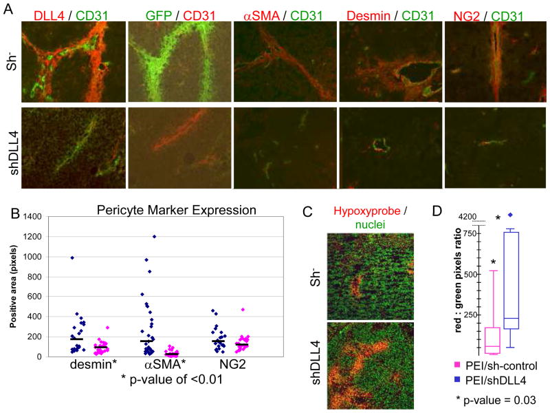
Figure 4.
Inhibition of DLL4 expression results in decreased tumor vessel-associated GFP+ BM-derived cells and pericytes, and increased hypoxia. A, B, TC71 cells were implanted subcutaneously on both flanks of nude mice that had previously been transplanted with GFP+ BM cells. Tumors were injected with PEI/sh− control (left side tumors) or PEI/shDLL4 (right side). A, Slides were stained with anti-CD31 in combination with anti-DLL4, anti-GFP, anti-α-SMA, anti-desmin, or anti-NG2. Colors are indicated above each panel. B, Five 10x fields from each tumor were analyzed using SimplePCI software. Diamonds represent the average positive area per slide for desmin, α-SMA, and NG2. Black bars show median values. Desmin, P < 0.01; α-SMA, P < 0.001; NG2, P not significant. C,D, Mice bearing bilateral TC71 tumors and treated with intratumor injections of PEI/sh− or PEI/shDLL4, described above, were injected with Hypoxyprobe-1 prior to sacrifice. Tumor sections were stained with anti-hypoxyprobe (red) to identify hypoxic regions and Sytox green (green) to identify nuclei. D, Five 10x fields from each tumor were analyzed. Box and whisker plots show the amount of hypoxia in PEI/sh− (pink) and PEI/shDLL4 (blue) treated tumors. P = 0.03.

