
Figure 7.
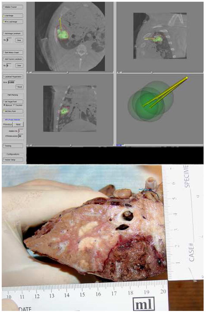
A four quadrant graphical user interface shows a composite treatment plan that includes four spherical ablations to cover the tumor. The planned treatment margins extend to the pleural surface because of the peripheral position of the tumor (top left). Likewise, a cross section of the explanted lung shows the extension of the coagulation to the pleural surface (bottom) suggesting correct anatomic correlation with the planned treatment.