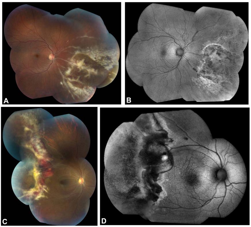
Figure 1.
Fundus photographs and corresponding FAF images of new-onset CMV retinitis in two HIV-positive patients. Fundus photograph of a 32 year-old male patient shows full-thickness retinitis in the inferonasal quadrant of the right eye (A). The FAF image demonstrated hyperautofluorescence at the posterior border of the zone of retinitis with mottled hyper- and hypoautofluorescence anterior to the advancing border in areas of clinically atrophic RPE (B). In a 35 year-old female patient with HIV and CNS lymphoma, a hemorrhagic retinitis is seen (C) and FAF imaging shows hyperautofluorescence at the posterior border of the active retinitis with a swath of hypoautofluorescence corresponding to hemorrhage, retinal edema, and full-thickness retinitis (D). Mottled hyper- and hypoautofluorescence are seen anterior to this area of active disease, corresponding to RPE atrophy seen in the fundus photograph.

