Figure 6.
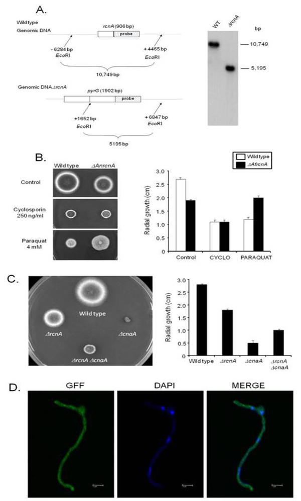
Molecular characterization of the A. nidulans AnrcnA gene. (A) Schematic illustration of the AnrcnA deletion strategy. (A) Genomic DNA from both wild type and ΔAnrcnA strains was isolated and cleaved with the enzyme EcoRI; a 2.0-kb DNA fragment from the 3'-noncoding region was used as a hybridization probe. This fragment recognizes a single DNA band (about 10.7-kb) in the wild type strain and also a single DNA band (about 5.2-kb) in the ΔAnrcnA mutant as shown in the Southern blot analysis. (B) Wild type and ΔAnrcnA mutant strains were grown for 72 hours at 37°C in complete medium in the absence or presence of cyclosporine A 250 ng/ml and paraquat 4 mM. (C) Growth phenotypes of A. nidulans wild type, ΔAnrcnA, ΔAncnaA, ΔAncnaA mutant strains were grown in complete medium for 72 hours at 37°C. In (B) and (C) graphs show the radial growth (cm) of the strains under different growth conditions. The results are the means ± standard deviation of four sets of experiments. (D) GFP::AnRcnA localizes to the cytoplasm. Germlings of the GFP::AnRcnA were grown in liquid MM+ 2% glycerol for 24 hs at 30°C. The germlings were treated or not with 50 mM calcium chloride for different periods of time from 5 to 60 minutes. After the treatment, germlings were analysed by laser scanning confocal microscopy. The figure shows a GFP::AnRcnA germling exposed to calcium chloride; however, germlings not exposed to calcium chloride displayed essentially the same results. Images were captured by direct acquisition. Bars, 5 μm.

