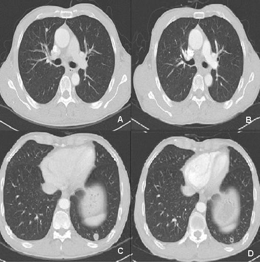
Figure 2.

A, B: Chest CT demonstrating hematogenous metastastatic nodule in RML (arrow, A) disappeared after 4th FOLFOX4 cycles (B). C, D: Chest CT demonstrating hematogenous metastastatic nodule in LLL (arrow, C) that formed fibrotic cavity after 4th FOLFOX4 cycles (D).
