
Fig 3.
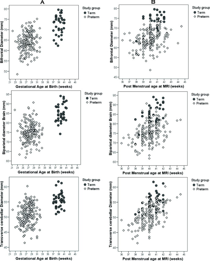
A and B, Comparison of bifrontal, biparietal, and transverse cerebellar diameters in preterm infants (diamond) and full-term infants (circle) by their gestational age at birth (A) and by their postmenstrual age at the time of MR imaging (B), demonstrating the lower values obtained in the preterm cohort.