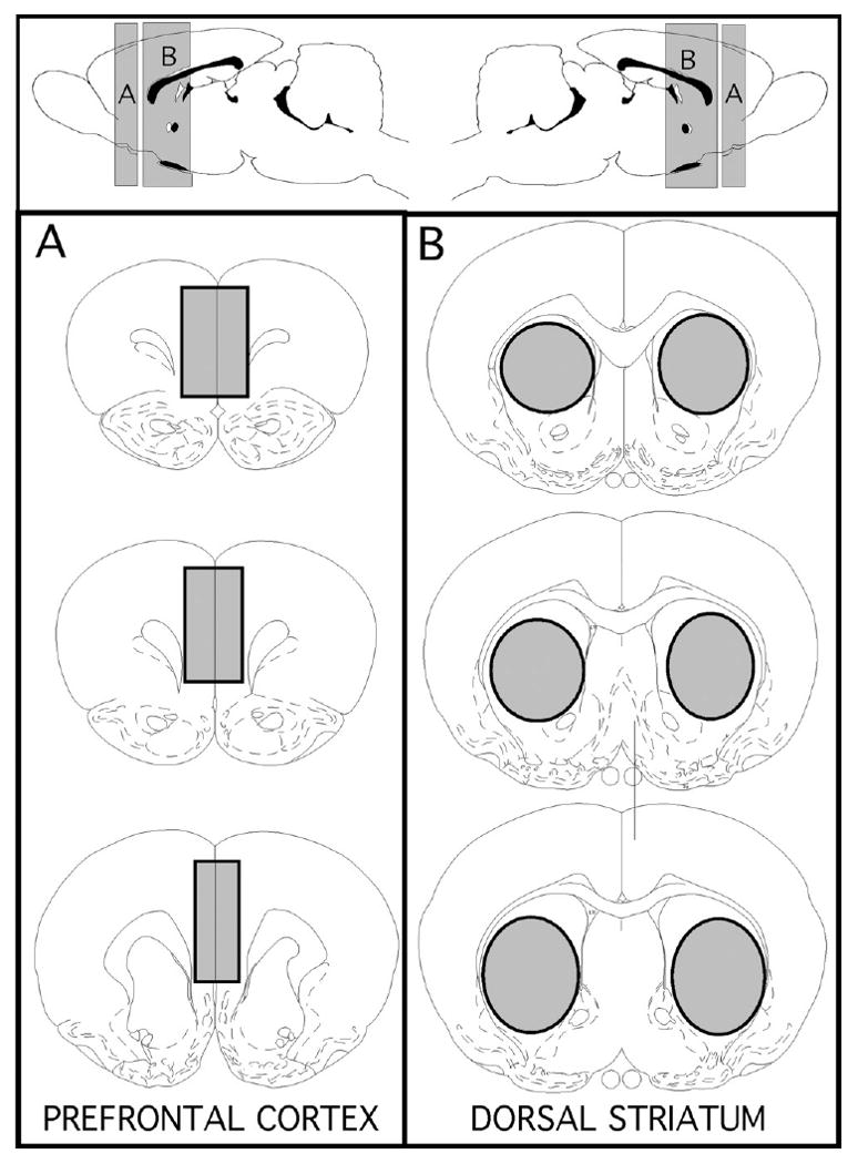
Fig. 1.

Schematic diagrams showing the anterior/poster (top panel) and coronal (lower left and right panels) locations of brain regions marked in grey that were micro-dissected out for the use in homogenate binding assays. The regions of interest that were collected were the pregenual medial prefrontal cortex (A) and dorsal striatum (B) taken at levels at and posterior to the anterior commisure. The templates showing these locations are modified from the atlas of Paxinos and Watson (1998).
