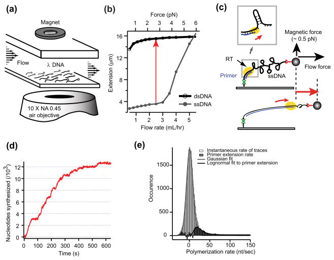
Figure 1. DNA replication on flow-stretched ssDNA by HIV-1 RT.
(a) Schematic diagram of the experimental setup. (b) Force-extension curves for ssDNA and dsDNA. The polymerase activity of ssDNA conversion to dsDNA can be monitored as a change in the extension length of each DNA molecule at a given force (red arrow). (c) Schematic of DNA polymerization by HIV-1 RT resulting in a lengthening of a ssDNA tether. (Inset) As HIV-1 RT encounters hairpin duplex during DNA polymerization, what is the mechanism for the replication on this region? (d) Example trajectory from tracking a DNA-tethered bead over time. (e) Histogram of HIV-1 RT instantaneous polymerization rates from raw polymerization trajectories at a stretching force of 3.0 pN. Effective plateaus are reflected by a Gaussian peak at around 0 nt/s. Original distribution subtracted with the Gaussian fit yields a population for the primer extension activity, which is fitted with a lognormal function (black line).

