Fig. 5.

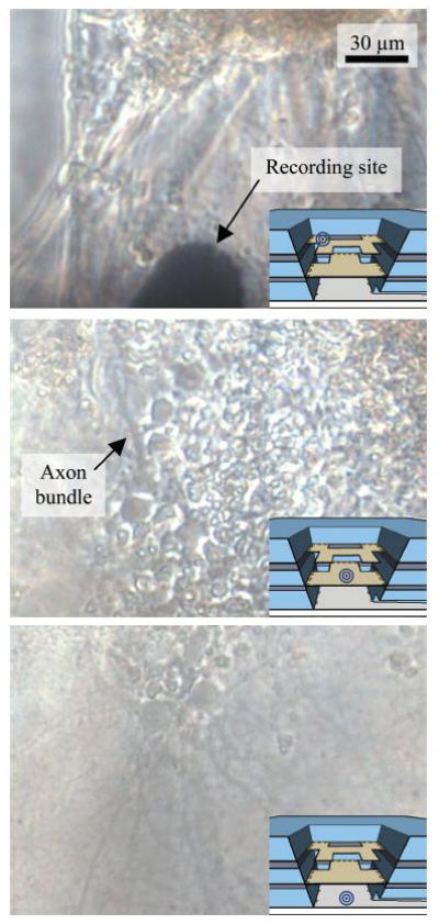
Micrographs of the three layers of the device with cells cultured at 18 DIV. The insets are cross-section of the cell chamber and show the imaging location for each micrograph. The scale is the same for all three images.

Micrographs of the three layers of the device with cells cultured at 18 DIV. The insets are cross-section of the cell chamber and show the imaging location for each micrograph. The scale is the same for all three images.