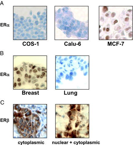
Figure 6.
Immunohistochemical analysis in NSCLC biopsy samples. A, Staining of paraffin-embedded COS-1, Calu-6, and MCF-7 cell pellets with ERα antibodies. B, Examples of breast and NSCLC tissue stained with ERα antibodies. C, A representative of lung tissue showing ERβ staining in cytoplasm only (left), and staining in both nucleus and cytoplasm (right). ERβ antibodies were from Upstate Biotechnology.

