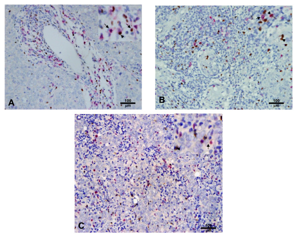
Figure 1.
Double immunohistochemical staining for CD8/Foxp3, Foxp3/GrB and Foxp3/IL-17. A. NPC tumor tissue with numerous CD8+ (red) and Foxp3+ (brown) cells (× 200). B. NPC tumor tissue with numerous Foxp3+ (brown) and GrB+ (red) cells (× 200). C. NPC tumor tissue with numerous Foxp3+ (red) and IL-17+ (brown) cells (× 200). The staining patterns show that CD8 is on the cell membrane, GrB and IL-17 are in the cytoplasm, and Foxp3 is in the nucleus. There are a few CD8+Foxp3+ and Foxp3+GrB+ cells, but no Foxp3+IL-17+ cells.

