Figure 3.
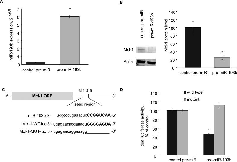
A: Hep-SWX cells were transfected with control-pre-miR and pre-miR-193b at 100 nM. After 72 hours, the expression of miR-193b was quantitated by real time PCR. Error bars indicate SEM.* p < 0.001 (n=3). B: Immunoblot analysis was performed in transfected Hep-SWX cells. Representative blots are shown along with quantitative data. Error bars indicate SEM (n=3). The relative expression of Mcl-1 to actin is expressed as a percentage of that in Hep-SWX cells. * p < 0.001. C: The location of target sites of miR-193b in the Mcl-1 3'-UTR are shown. The sequence of the mutated Mcl-1 3’-UTR construct with mutations to delete the putative miR-193b binding site is also displayed. D: Hep-SWX cells were transfected with the Renilla luciferase expression construct pRL-tk, the luciferase construct Mcl-1-WT-luc or Mcl-1-MUT-luc (in which the Mcl-1 target site was deleted) and miR-193b or control precursor. The data represent mean and SEM from 3 determinations from four independent transfections. * p < 0.001 compared to the respective controls.

