Figure 1.

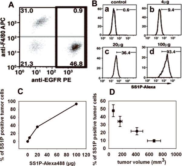
In vivo tumor cell staining by SS1P-Alexa. A, Cell composition of tumor cell suspension. Tumor cell suspension prepared by digestion was stained by anti-EGFR PE and anti-F4/80 APC to distinguish A431/H9 cells and mouse macrophages. B, The staining of A431/H9 tumors. Control mice received saline (Ba). Mice bearing A431/H9 tumors received SS1P-Alexa488 of different doses, 4 μg (Bb), 20 μg (Bc) and 100 μg (Bd). After 3 h, the staining of SS1P-Alexa on tumor cells (gated population in A) was analyzed. Numbers denote the percentage of SS1P-Alexa positive tumor cells as total of tumor cells. C, Summary of tumor staining by increased doses of SS1P-Alexa. Data from Ba, Bb, Bc and Bd were plotted. D, The effect of tumor size on tumor cell staining. Mice bearing A431/H9 tumors of various size (70, 170, 400 and 650 mm3) received 20 μg of SS1P-Alexa. Tumor cell staining after 3 h was measured (n = 5).
