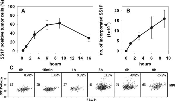
Figure 3.
In vivo time course for internalized SS1P in tumor cells. SS1P(E553D)-Alexa (20 μg) was given to mice bearing A431/H9 tumors. Tumors were removed at different time points, 15 min, 1, 3, 6, 9 and 16 h (n = 3). The staining of tumor cells by SS1P(E553D)-Alexa was analyzed. The percentage of SS1P(E553D)-Alexa positive tumor cells was shown in A. The number of SS1P-Alexa molecules incorporated into tumor cells was shown in B. A representative result from each time point was shown in C. The number at the upper right corner of each time point denotes the percentage of SS1P(E553D)-Alexa positive tumor cells. The number in the left above the middle line is the MFI of tumor cell population.

