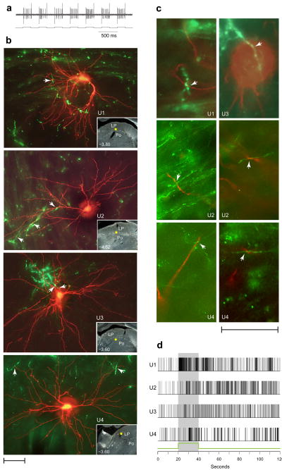
Figure 3.
Close apposition between dura/light-sensitive neurons and retinal afferents in LP and Po. (a) Synchronization of neuronal activity (top trace) with the current (bottom trace) delivered by the TMR–dextran filled recording micropipette (see text for detail). (b) Dura/light sensitive units (U1–U4) filled with TMR–dextran (red) and retinal axons labeled anterogradely with CTB (green). Each image represents z-stacking of approximately thirty 1–1.5-μm-thick scans. Arrowheads point to potential axodendritic or axosomatic apposition. Localization of each cell body is marked by a yellow star in the low-power, darkfield inset. Numbers indicate distance from Bregma. (c) Evidence for axodendritic and axosomatic apposition within a single 1–1.5-μm-thick scan taken from the units shown in b. (d) Neuronal firing in response to 50,000 lux of white light (green line and shaded area), corresponding to the individual neurons shown in b. Scale bars represent 50 μm (b,c).

