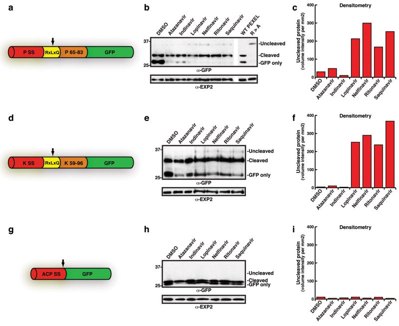
Figure 1. PEXEL processing is sensitive to HIV protease inhibitors.
a, Structure of PfEMP3-GFP. PSS, signal sequence; RxLxQ PEXEL; residues 65-83 of PfEMP3. Arrow, PEXEL cleavage. b, PfEMP3-GFP processing with HIV-1 inhibitors. c, Densitometry of uncleaved bands in b. d, Structure of KAHRP-GFP. KSS, signal sequence; RxLxQ PEXEL, residues 59-96 of KAHRP. e, KAHRP-GFP PEXEL processing with inhibitors. f, Densitometry of uncleaved bands in e. g, Structure of ACPs-GFP. ACP SS, signal sequence-GFP. Arrow, signal sequence cleavage. h, ACPs-GFP signal sequence processing is not inhibited. ‘Cleaved’, signal sequence cleavage. i, Densitometry of the uncleaved region of immunoblot. (see Supplementary for details)

