Fig. 4.

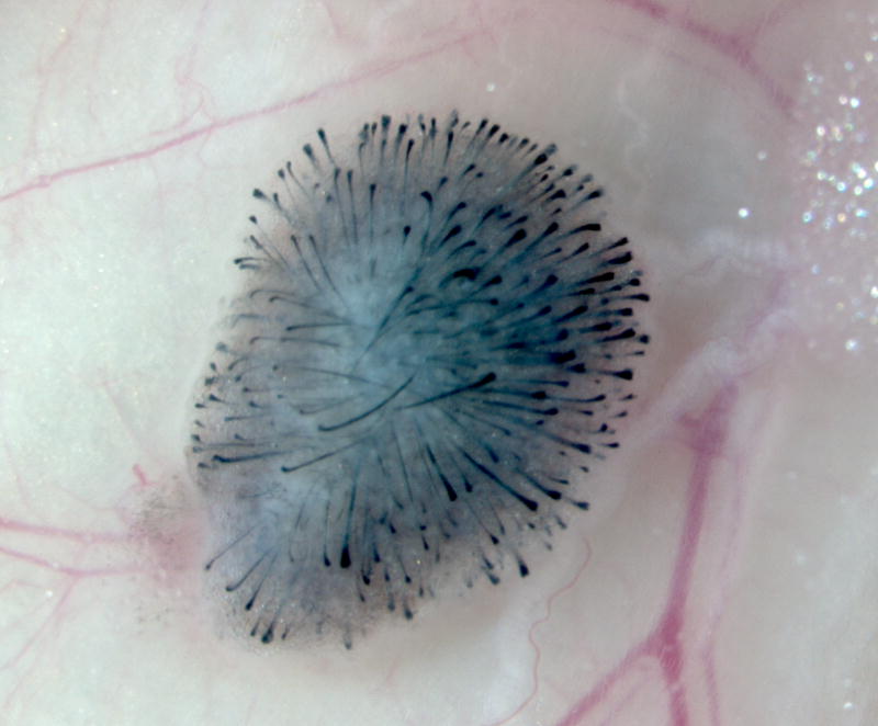
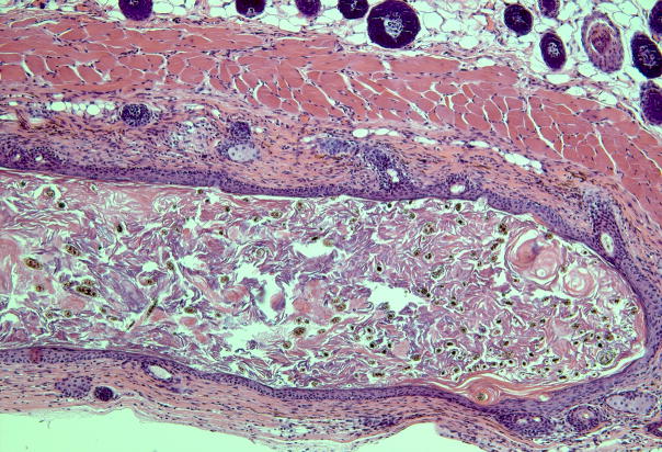
Subcutaneous injection of dissociated murine epidermal and dermal cells into the back skin of a nude mouse. (a) A nodule or “patch” with regenerated hair follicles can be observed from the undersurface of the skin in 2 weeks. (b) The histology shows formation of a cyst with hair follicles coming out from the cyst wall.

