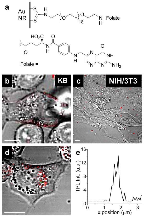
Figure 6. In vitro cellular imaging with F-NRs (34).
(a) folate-oligoethyleneglycol ligands, conjugated onto NR surface by in situ dithiocarbamate formation. (b) F-NRs bound to the KB cell surface after incubation for 6 hours. (c) Almost no F-NRs were observed to be associated with NIH/3T3 cells, which do not overexpress the high-affinity folate receptor. (d) F-NRs were internalized into KB cells and delivered to the perinuclear area after incubation for 17 hours. (e) TPL intensity profile across green line in (d). Bar= 10 μm.

