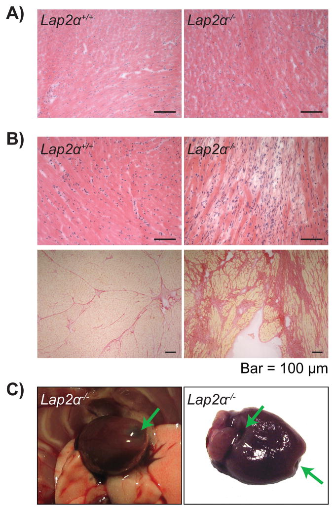
Figure 3. Old Lap2α−/− mice show sporadic cases of fibrosis.
A) Young male Lap2α−/− myocardium is histologically indistinguishable from the WT. B) 18% of the old male Lap2α−/− hearts show disperse fibrotic foci (upper panels haematoxylin & eosin-stained, lower panels Picrosirius Red-stained heart sections). C) Thinning of the old Lap2α−/− myocardium – arrows mark the transparent fibrotic regions of the heart.

