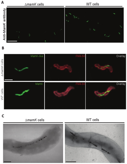
Figure 7. Detection of MamK and MamK-like filaments in fixed cells of M. magneticum AMB-1 WT and ΔmamK strains.
The primary antibody was the anti-MamK antibody and the secondary antibody was coupled to FITC. Confocal microscope data acquisition parameters: 3D stacks of 2048 x 2048 pixel images, 0.16 µm steps, 4x frames average, 2x line accumulation. A) FITC fluorescence emission at λ = 518 nm (excitation at λ = 488 nm), 2.4x microscope zoom. Left panel, ΔmamK mutant cells; right panel, WT cells. Scale bar: 10 µm. B) Cell membranes were stained with FM® 4-64 FX. Upper panels, ΔmamK mutant cells; lower panels, WT cells. Left column, FITC fluorescence emission at λ = 518 nm (excitation at λ = 488 nm); middle column, FM® 4-64 FX fluorescence emission at λ = 744 nm (excitation at λ = 633 nm); right column, left and middle column images overlaid; 4.74x microscope zoom for all panels. Scale bar: 1 µm. C) Visualization of magnetosome alignment with TEM. Left panel, ΔmamK cells (unstained); right panel, WT cells (1% uranyl acetate stained). Scale bar: 300 nm.

