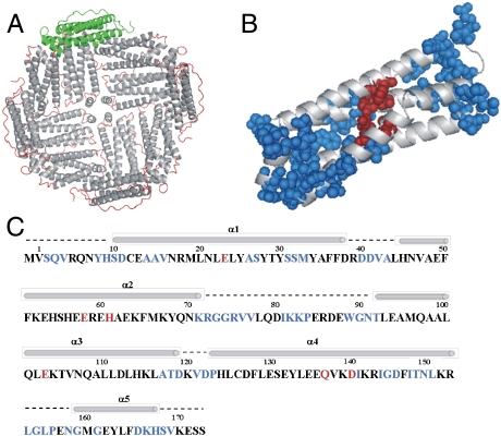
Fig. 1.
Sequential assignment of ferritin by solid-state NMR. (A) 3D structure of recombinant frog (R. catesbeiana) M ferritin (24 subunits, PDB 1MFR; PyMOL 0.99rc6). The four-helix-bundle subunits are displayed as gray helices and red loops. The green subunit illustrates the orientation of the monomeric units along the surface of the hollow sphere. (B) Representation of the four-helix-bundle subunit NMR-assigned residues (blue spheres) and ferroxidase residues (red spheres). (C) Primary and secondary structures of frog M ferritin-ferroxidase site residues (red font), NMR sequence-specific assigned residues (blue font), α-helices (bars), and loops (broken lines).

