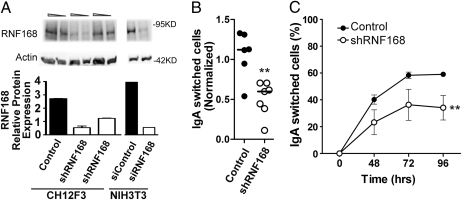
Fig. 3.
CSR is impaired in RNF168-deficient CH12F3-2 cells (A) Western blot analysis of control or shRNF168 CH12F3-2 cells quantifying RNF168, with β-actin as the loading control. NIH 3T3 cells treated with control siRNA and siRNF168 were used as negative and positive controls for RNF168 protein expression, respectively. (B) Six individual control and seven individual shRNF168 CH12F3-2 clones were stimulated for 2 days and then analyzed for IgA expression. Values were normalized by dividing the % IgA-positive cells in the experimental group into the % IgA-positive cells in the stimulated parental CH12F3-2 cells. CSR assays were performed on each clone in triplicate, and statistical significance was evaluated by the two-tailed t test. **P = .0023 (C) Three control and three shRNF168 CH12F3-2 clones were stimulated for 2, 3, and 4 days, after which IgA expression was analyzed. CSR assays were performed in duplicate for each clone. Statistical significance was tested by two-way ANOVA. **P = .0034.

