Fig. 2.
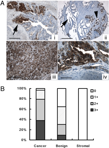
Binding of mAb F77 to prostate tissue samples. (A) mAb F77 immunohistochemical staining in prostate tissue. In benign prostate glands (i) there is a mosaic staining pattern of some benign prostate glands. Arrow points to the F77-positive prostate gland. (Scale bar, 100 μm.) Benign prostate glands are negative for mAb F77 (arrow, ii), whereas cancerous prostate glands are positive (arrowhead, ii). (Scale bar, 200 μm.) In high-grade prostate cancers (iii) poorly differentiated prostate cancerous tissues are diffusely positive for mAb F77. (Scale bar, 50 μm.) In bone metastasis (iv) cancerous glands in a bone metastasis are also diffusely positive. (Scale bar, 25 μm.) (B) Quantification of Immunohistochemical staining of mAb F77. Specimens (n = 116) were stained with mAb F77 (1 μg/mL). Staining intensity of the tissues was graded as 0 (negative), 1+ (weak), 2+ (moderate), and 3+ (strong).

