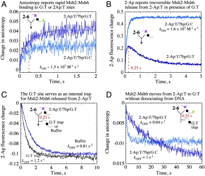
Fig. 3.
Msh2-Msh6 moves from 2-Ap:T to G:T without dissociating from DNA. (A) Fluorescence anisotropy shows Msh2-Msh6 binding mismatched 2-Ap:T/7 bp/G:TTAMRA and matched 2-Ap:T/7 bp/G:CTAMRA DNA rapidly (kON = 1.35 and 1.4 × 10⁷ M-1 s-1, respectively). (B) 2-Ap fluorescence yields the same rate for 2-Ap:T/7 bp/G:CTAMRA, but 2-Ap:T/7 bp/G:TTAMRA shows biphasic kinetics. (C) 2-Ap fluorescence reports rapid release of Msh2-Msh6 from 2-Ap:T in 2-Ap:T/7 bp/G:TTAMRA with an external trap (unlabeled G:T) or with only buffer added after 0.25 s (kOFF = 0.8–1.2 s-1). (D) Corresponding TAMRA anisotropy shows slow release of Msh2-Msh6 from 2-Ap:T/7 bp/G:TTAMRA (kOFF = 0.044 s-1).

