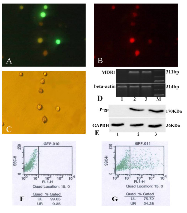
Figure 1.
BMCs infected with Ad-EGFP-mdr1. Daunomycin efflux assay detected by fluorescence microscope. A: BMCs incubated with Ad-EGFP-MDR1 for 48 h expressed EGFP(green). × 400 B: Daunomycin (red) aggregated inside the BMCs which were not infected with the adenovirus. × 400 C: Bright field images of those BMCs × 400. MDR1 mRNA in BMCs was detected by RT-PCR. D: The expected size band of human MDR1 mRNA was 311 bp. The expected size band of mouse beta-actin was 314 bp. The expression of P-gp in BMCs was assessed by western blot. E: Ad-EGFP-mdr1 infection induces expression and production of human P-gp in BMCs. Flow cytometry determined percentage of green fluorescence. BMCs infected with Ad-EGFP-mdr1 successfully would show green under fluorescence channel analyses. F: the background was about 0.4%. G: The infection rate of BMCs incubated with Ad-EGFP-mdr1 for 48 h was about 24.3%, 1.BMCs. 2.BMCs incubated with Ad-EGFP-mdr1 for 48 h. 3. Positive control. M:marker.

