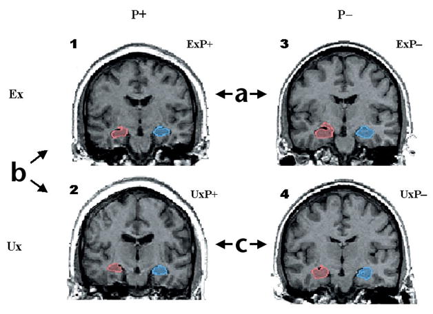
Fig. 1.

Discordant monozygotic twin paradigm for assessing MRI differences in PTSD. Sample coronal MRI images of right (red) and left (blue) hippocampi in a PTSD and a non-PTSD twin pair. Images represent four subject groups: (1) combat-exposed (Ex) subjects who developed chronic PTSD (ExP+); (2) their combat-unexposed (Ux) co-twins with no PTSD themselves (UxP+); (3) Ex subjects who never developed PTSD (ExP−) and (4) Ux co-twins also with no PTSD (UxP−). Contrast (a) provides a replication of previous work demonstrating smaller hippocampal volumes in combat veterans with versus without PTSD. Contrast (b) identifies the neurotoxicity effect—hippocampal reduction—as environmentally acquired, by contrasting hippocampal volumes in combat-exposed PTSD veterans with their unexposed co-twins. Contrast (c) examines pre-existing vulnerability by contrasting hippocampal volumes in the two groups of combat-unexposed co-twins whose combat-exposed brothers did versus did not develop PTSD. Model is tested by a diagnosis (P+ versus P−) × exposure (Ex versus Ux) ANOVA. Diagnosis refers to combat-exposed twin only. If hippocampal volume represents a vulnerability factor, the model predicts a significant main effect of diagnosis in the absence of a diagnosis × exposure interaction (that is, PTSD combat-exposed veterans and their unexposed co-twins show the same pattern). If hippocampal reduction results from neurotoxicity, the model predicts a significant main effect of exposure and/or a significant diagnosis × exposure interaction.
