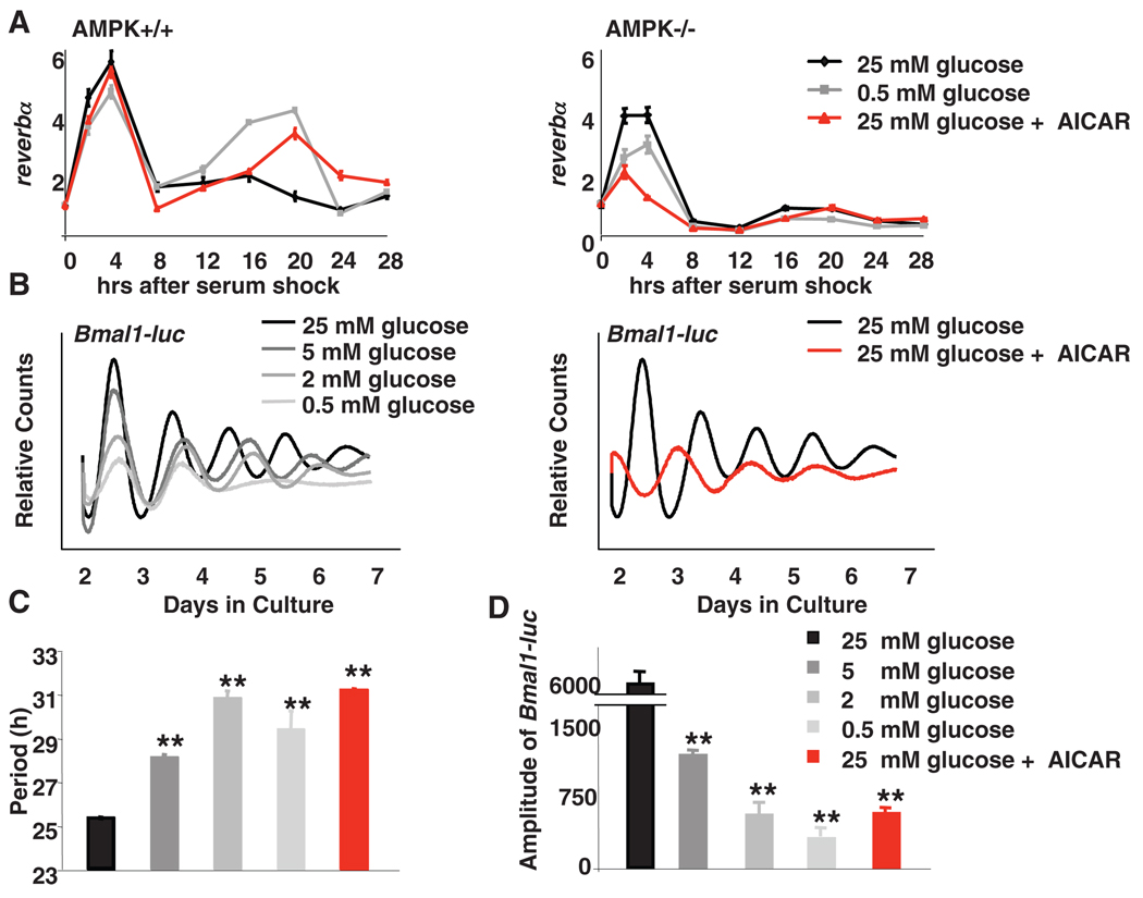
Fig. 2.
Disruption of AMPK signaling alters circadian rhythms in MEFs. (A) MEFs were synchronized by serum shock and transferred to media containing glucose and AICAR as indicated. Quantitative PCR (QPCR) was performed by using cDNA from samples collected at the indicated times. Data are means ± SD of a representative experiment analyzed in triplicate. (B) Typical results of continuous monitoring of luciferase activity from U2OS cells stably expressing Bmal1-luciferase. (C and D) Quantification of the circadian period (C) and amplitude (D) of Bmal1-driven luciferase from experiments performed as described in (B). Data in (C) and (D) are means ± SD for four samples per condition. Analysis of variance (ANOVA) indicated a significant difference between categories. **P < 0.01 versus samples cultured in 25 mM glucose.

