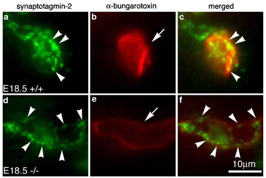
Figure 2.
Differentiation of the NMJ in the absence of the γ-subunit. High magnification views of a single endplate from wholemount diaphragm muscle at E18.5, double-labeled with synaptotagmin-2 (Syt-2) antibody (green) and Texas-red conjugated α-bungarotoxin (red). Individual endplate (AChR cluster) in the mutant (e, arrow) appeared less intensely labeled by α-bungarotoxin, but their sizes are bigger, compared to the wild type (b, arrow). In both WT and mutant muscles, Syt-2 antibody staining was highly concentrated at the nerve terminals (arrowheads in a, d). Scale bar 10 µm

