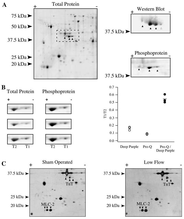
Fig. 3.

Phosphorylation of Troponin T and MLC-2. a Total protein stain of a homogenate resolved by 2D SDS-PAGE using a 3-5.6NL strip. The putative TnT isoelectric variants are marked (left). Western blotting using a TnT antibody (right, top) or phosphoprotein staining (right, bottom) were carried out to identify phosphorylated (▲) or nonphosphorylated (△) forms of TnT. b Panels focusing on the two predominant TnT isoelectric variants of the more abundant lower Mr isoform, marked T1 and T2, stained in triplicate by total protein stain and phosphoprotein stain (left). T1/T2 ratio for the total protein and phosphoprotein stain, as well as the quotient of the phosphoprotein T1/T2 value over the total protein T1/T2 value (right). c Two-dimensional SDS–PAGE of representative total homogenates from a sham operated and low flow heart, identifying TnT and MLC-2 isoelectric variants
