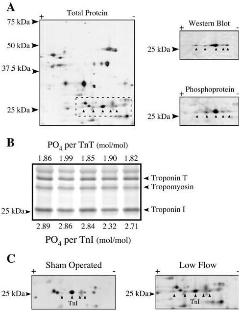
Fig. 5.

Phosphorylation of Troponin I. a Total protein stain of a homogenate resolved by 2D SDS–PAGE using the cathodic half of a 7–11NL strip. The putative TnI isoelectric variants are marked (left). Western blotting using a TnI antibody (right, top) or phosphoprotein staining (right, bottom) were carried out to identify the TnI variants, all of which were phosphorylated. b Five rat heart homogenates with known amounts of TnT phosphorylation were resolved by SDS–PAGE and stained with Pro-Q Diamond phosphoprotein stain. The TnI phosphoprotein signal from each sample was measured and the stoichiometry determined based on a comparison to the TnT phosphoprotein signal. c Two-dimensional SDS–PAGE of representative sham operated and low flow rat heart homogenates stained for total protein, identifying the isoelectric variants of TnI
