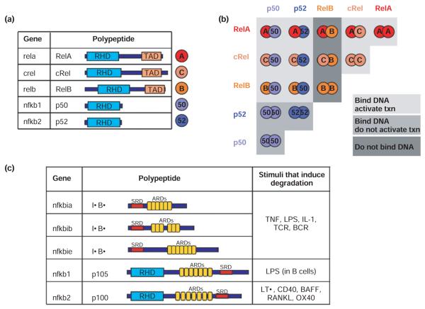
FIGURE 1.
The components of the NF-κB signaling system a) The NF-κB family members. The gene name is indicated for each polypeptide. Each NF-κB family member has a Rel Homology Domain (RHD) for dimerization and DNA binding. RelA, cRel, and RelB have Transcriptional Activation Domains (TAD). b) The 5 NF-κB monomers can combine to form 15 potential dimers. Of these, 9 can bind DNA and activate gene transcription (light grey), 3 (the p50 or p52 only containing dimers) bind DNA but do not activate transcription (medium grey), and 3 do not bind DNA (dark grey). c) The IκB protein family members and signals that induce the degradation of each. The ARDs on p105 and p100 (which are proteolytically processed to p50 and p52 NF-κB monomers, respectively) can act to self-inhibit p50 and p52. p100 can form a multimeric complex in which it can inhibit other latent NF-κB dimers. BCR = B cell receptor; TCR = T cell receptor RHD = Rel Homology Domain; ARD = ankyrin repeat domain; TAD = transcriptional activation domain, SRD = signal response domain.

