
Figure 1. The structure of a CRISPR locus.
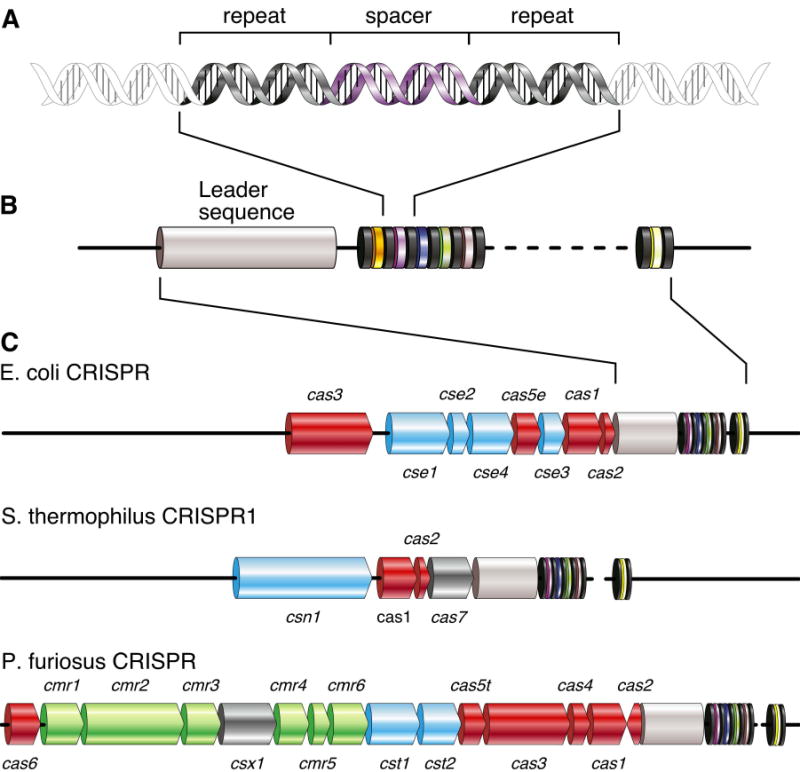
(A) Repeat sequences averaging 32bp are interleaved by variable spacers of approximately the same size. (B) The number of repeat-spacer units varies greatly. A conserved leader sequence (gray) of several hundred base pairs is located on one side of the cluster. (C) CRISPR-associated (cas) genes surround the CRISPR locus. Three examples of well-studied CRISPR loci are shown. Core cas genes are depicted in red, subtype-specific genes in blue, and the RAMP module in green. Unclassified genes are shown in dark grey. Gene names follow the nomenclature of Haft et al., except cas7, which was named by Barrangou et al.