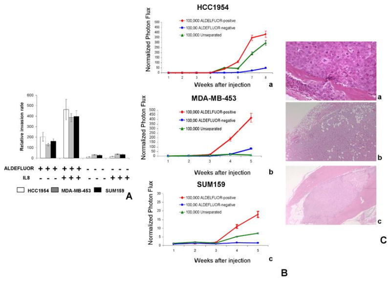
Fig. 5. ALDEFLUOR-positive cells display increased metastatic potential.

A. The IL8/CXCR1 axis is involved in cancer stem cell invasion. The role of the IL8/CXCR1 axis in invasion was assessed by a Matrigel invasion assay using serum or IL8 as attractant for three different cell lines (HCC1954, MDA-MB-453, SUM159). ALDEFLUOR-positive cells were 6- to 20-fold more invasive than ALDEFLUOR-negative cells (p<0.01). When using IL8 (100 ng/ml) as attractant, we observed a significant increase of ALDEFLUOR-positive cells invading through Matrigel compared to serum as attractant (p<0.05). In contrast IL8 had no effect on the invasive capacity of the ALDEFLUOR-negative population. B-C. The ALDEFLUOR-positive population displayed increased metastatic potential. Ba-c. Quantification of the normalized photon flux measured at weekly intervals following inoculation of 100,000 luciferase infected cells from each group (ALDEFLUOR-positive, ALDEFLUOR-negative, unseparated). Mice inoculated with ALDEFLUOR-positive cells developed several metastasis localized at different sites (bone, muscle, lung, soft tissue) and displayed a higher photon flux emission than mice inoculated with unseparated cells, which developed no more than one metastasis per mouse. In contrast, mice inoculated with ALDEFLUOR-negative cells developed only an occasional small metastasis, which was limited to lymph nodes. C. Histologic confirmation, by H&E staining, of metastasis in bone (Ca), soft tissue (Cb) and muscle (Cc) resulting from injection of ALDEFLUOR-positive cells.
