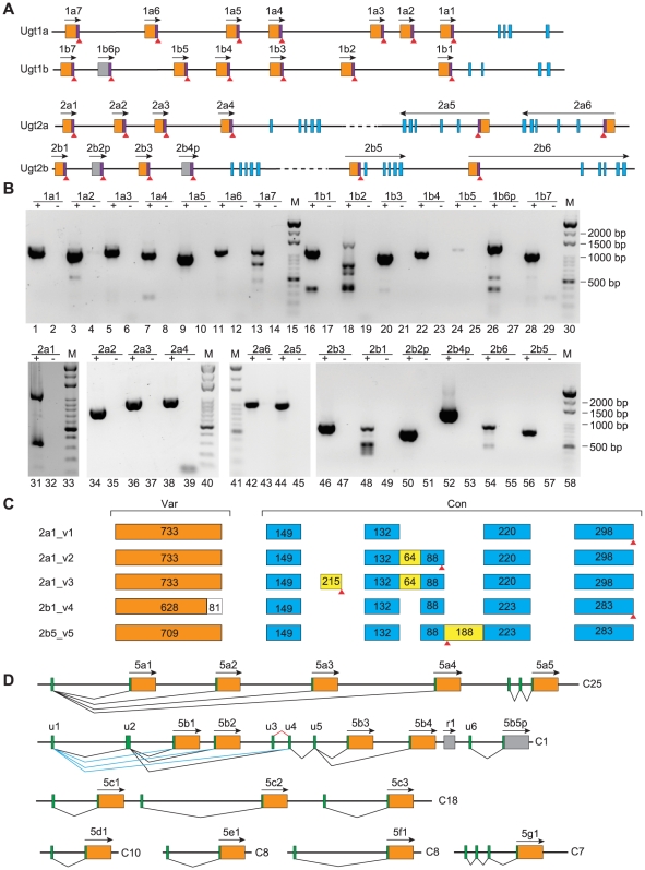
Figure 2. Alternative splicing of the zebrafish Ugt genes.
(A) Each Ugt1 or Ugt2 member has a short-form transcript which corresponds to the variable exon (orange box) and its immediate downstream intronic sequences (purple box). Red triangles indicate the potential translational stop codons. Gray box indicates pseudogene. Transcription directions are marked by an arrow above each gene. (B) Detection of the short-form cDNA by RT-PCR and agarose electrophoresis. “+” and “−” indicate with and without reverse transcriptase, respectively. Amplification bands are detected only in “+” lanes. M: 1 kb marker. (C) Alternatively spliced variants for the Ugt2a1, and Ugt 2b1 and 2b5 genes. The exon length is shown in each box. (D) Alternative splicing of the zebrafish Ugt5 genes. Green boxes represent 5′ noncoding exons or short noncoding exonic sequences immediate upstream of the ATG codon.

