
Figure 1.
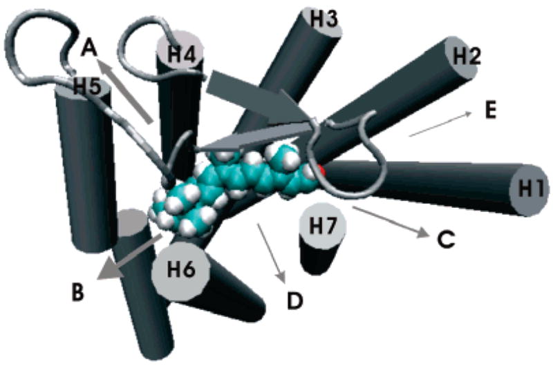
Retinal egress pathways near the extracellular side (out of the page toward the reader). They are classified into five (A, B, C, D, and E) pathways according to the helices around the egresses. The arrow widths represent the relative frequencies. See Figure 3 in Supporting Information for a stereoview.