
Fig. 4.
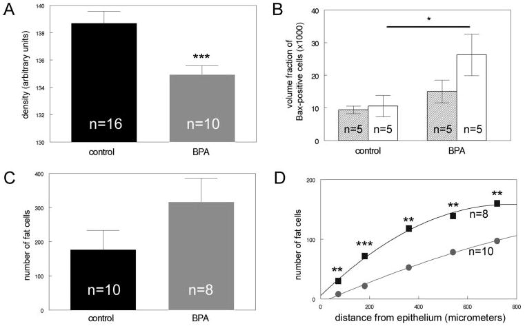
Alterations are observed in the developing fat pad of BPA-exposed females, compared with controls. A, The OD of the fat pad in BPA-exposed animals (gray) is decreased, compared with controls (black). B, Expression of the proapoptotic marker Bax is significantly increased in the developing fat pad (white) in BPA-exposed animals relative to controls. No statistically significant differences were noted in the loose connective tissue (shaded). C, The total number of cells containing fat vacuoles was increased in BPA-exposed females, compared with controls, although this increase was not significant. D, The number of adipocytes was significantly increased within 1 mm from the developing epithelium in BPA-exposed females (squares), compared with controls (circles). *, P < 0.05; **, P < 0.02; ***, P < 0.005.