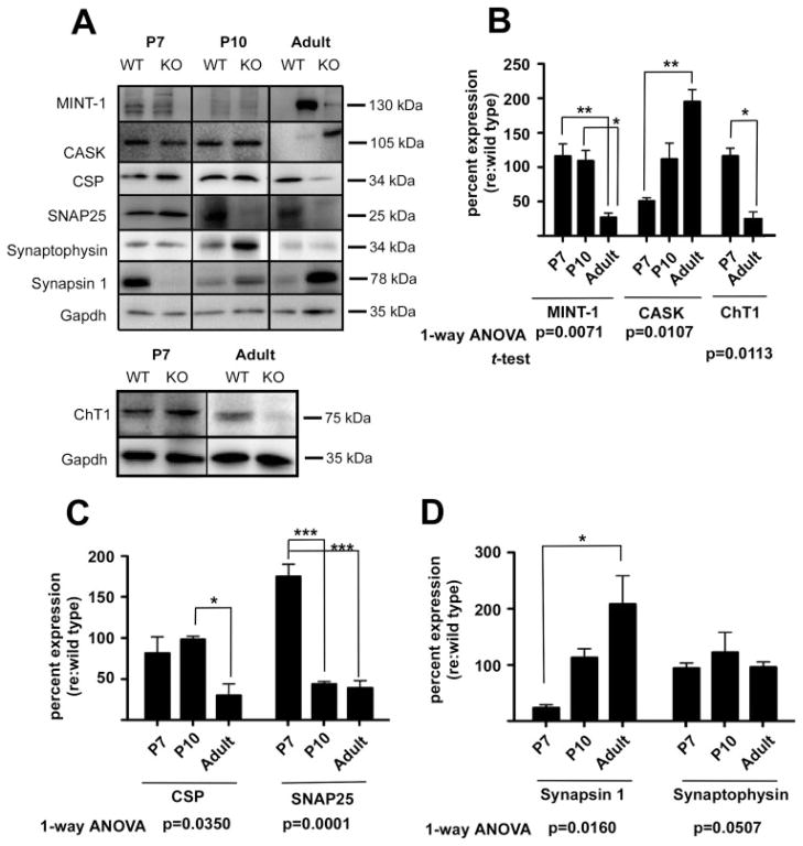
Figure 5.
Loss of α9 nAChR results in changes in presynaptic active zone proteins and synaptic vesicle binding proteins. (A) Total cochlear lysates from α9 null and wild-type control mice at P7, P10, and adult (2 months) ages were probed by western blotting with antibodies against trans-synaptic and signaling proteins as indicated. Western blots reveal that loss of α9 nAChR activity results in marked changes in expression of presynaptic SNARE proteins, scaffolding proteins and synaptic vesicle binding proteins in the α9 nulls at early postnatal and adult ages. Additionally, lysates from P7 and adult cochleae were probed to assess expression levels of the high affinity choline transporter ChT1. (B–D) Time series bar graphs show percent change in protein expression levels in α9 nulls relative to the wild type as discussed for Figure 4. These data demonstrate that α9 nAChR is required for the normal expression of presynaptic components that are essential for normal synapse assembly and function. Although synaptophysin levels remain essentially unchanged in the α9 null mice (D), the other proteins probed fall into distinct families of expression profiles. Thus, synapsin and CASK both begin expression at levels lower than that of wild type values and steadily increase in expression levels to adult ages (B,D). Mint-1, CSP, and ChT1 are normally expressed at early ages and then decrease to approximately 50% or less relative to wild type values by adult ages (B,C). SNAP-25 is the only protein probed that starts at higher than normal levels of expression (C). One-way ANOVA and Tukey multiple comparisons analyses revealed significant changes in expression over time in α9 null mice for all proteins examined except for synaptophysin, which was just outside of the significance margin (*p < 0.05; **p < 0.005; ***p < 0.0001).

