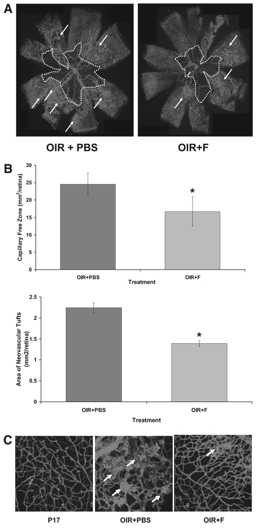
FIGURE 1.
(A) Flatmounted mouse retinas stained with isolectin B4 to identify areas of neovascularization (arrows) and capillary-free zones (dotted white line). (B) Histograms representing the results of morphometric analysis of retinal flatmounts measuring the capillary-free areas (upper bars) and neovascular tufts (lower bars). OIR+PBS, sham ischemic mouse retina at postnatal day 17; OIR+F, ischemic mouse retina at postnatal day 17 injected with 10 mg/kg/d fluvastatin (P12–P17). *P < 0.01 vs. OIR+PBS; x = mean ± SE; n = 11. (C) Three-dimensional images (z-stacks, 20× magnification) of retinal flatmounts stained with isolectin B4 (Texas Red demonstrating retinal capillary morphology in the different treatment groups).

