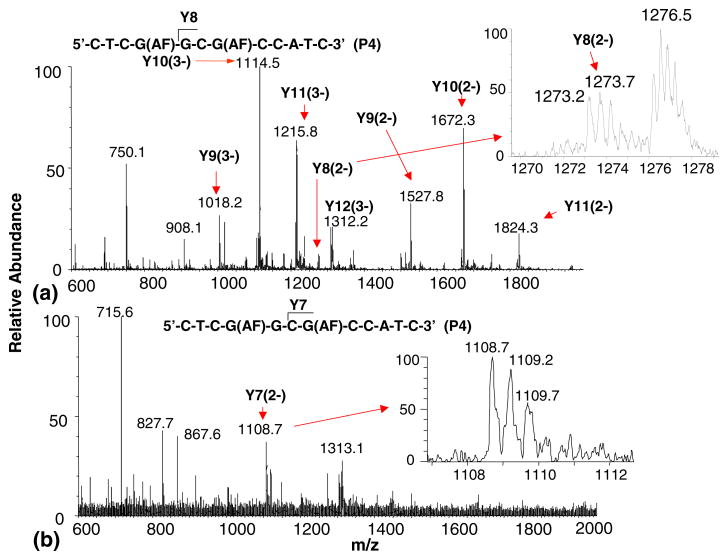
Figure 5.
ESI-TOF mass spectrum of (a) the hydrolysis fragments formed from the 5′-exonuclease digestion of product 4 after 80 min. The inset shows the Y8 fragment formed by the loss of a modified guanine nucleotide (spectrum summed over 3.5 to 7 min retention time); (b) of the hydrolysis fragments formed from the 5′-exonuclease digestion of product 4 after 80 min. The inset shows the Y7 fragment formed by the consecutive losses of a modified guanine (G1) and unmodified guanine (G2) nucleotide (spectrum summed over 2.5 to 3.5 min retention time).

